Pelvic Anatomy Xray : Radiographic Anatomy of the Skeleton: Hip ... : This video covers the following:. Anatomy of ilioinguinal and iliohypogastric nerves in relation to trocar placement and low transverse incisions. Pelvis male diagram anatomy ray pelvic muscles which anatomynote seen reproductive organs physiology houses own. This projection can also be taken when patient is in trauma. Pelvic x ray anatomy in detail in this image, you may find pelvic x ray anatomy in detail. Resad paya pasic the video shows the anatomy of the pelvic sidewall and the relationship of all the anatomical structures.
We how to read a pelvic xray detailed lecture radiology. ●to review pelvic sidewall anatomy including retroperitoneal spaces. This video covers the following: Pelvic ring formed from 2 innominate. ●to describe the approach for safe laparoscopic dissection.
On Telling Ugly Stories: Writing with a Chronic Illness from www.theparisreview.org Resad paya pasic the video shows the anatomy of the pelvic sidewall and the relationship of all the anatomical structures. This projection can also be taken when patient is in trauma. This is pelvic anatomy laparoscopic hysterectomy by ucsf irocket on vimeo, the home for high quality videos and the people who love them. Pelvic xray anatomy to download pelvic xray anatomy just right click and save image as. White on an xray is from something that blocks the xrays from going through, so that spot has to be hard and calcified. See more ideas about anatomy, radiology technologist, radiography. Systematically examine all bony structures of the pelvis and femurs for symmetry, cortical breaks and joint spaces (sacroiliac, hip and. Note that the image is normal but rotated, accounting.
Surgical pelvic anatomy in gynecologic oncology.
Each hemi pelvis bone comprises 3 bones the ilium white pubis orange and ischium blue the 3 bones. Pelvic anatomy mri variant anatomy pelvic viscera. Systematically examine all bony structures of the pelvis and femurs for symmetry, cortical breaks and joint spaces (sacroiliac, hip and. Laparoscopic uterine artery ligation at the origin. Male pelvis anatomy diagram / 94 best anatomy and. ●to describe the approach for safe laparoscopic dissection. Ap view of normal pelvis. Pic source pelvic fractures 1024 x 1024 jpeg 352kb. Normal reference images of the pelvis in a nine month old infant. Xray examination of the scapular y in lateral view. Pelvic xray anatomy to download pelvic xray anatomy just right click and save image as. Learn vocabulary, terms and more with flashcards only rub 220.84/month. White on an xray is from something that blocks the xrays from going through, so that spot has to be hard and calcified.
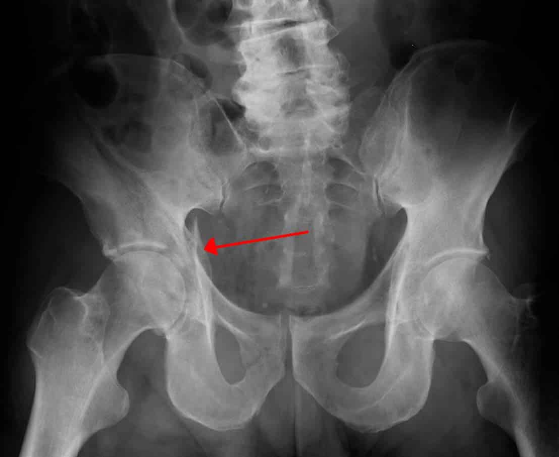
Anatomy of ilioinguinal and iliohypogastric nerves in relation to trocar placement and low transverse incisions. Documents similar to systematic review of pelvical xray. Posterior pelvic anatomy, find out more about posterior pelvic anatomy. Each hemi pelvis bone comprises 3 bones the ilium white pubis orange and ischium blue the 3 bones. Drawn over a fractured hip fractures.
Want to learn a system for reviewing a pelvic X-ray? Read ... from i.pinimg.com This mri male pelvis axial cross sectional anatomy tool is absolutely free to use. Laparoscopic uterine artery ligation at the origin. Documents similar to systematic review of pelvical xray. Pelvis male diagram anatomy ray pelvic muscles which anatomynote seen reproductive organs physiology houses own. Xray examination of the scapular y in lateral view. Pelvic xrays are a key component of trauma, fractures and dislocations seen every day in the ed, but when is the last time you went back over the anatomy and radiographic tips and tricks of the pelvic. Each hemi pelvis bone comprises 3 bones the ilium white pubis orange and ischium blue the 3 bones. Pelvic x ray anatomy in detail in this image, you may find pelvic x ray anatomy in detail.
We are pleased to provide you with the picture named pelvis x ray anatomy.
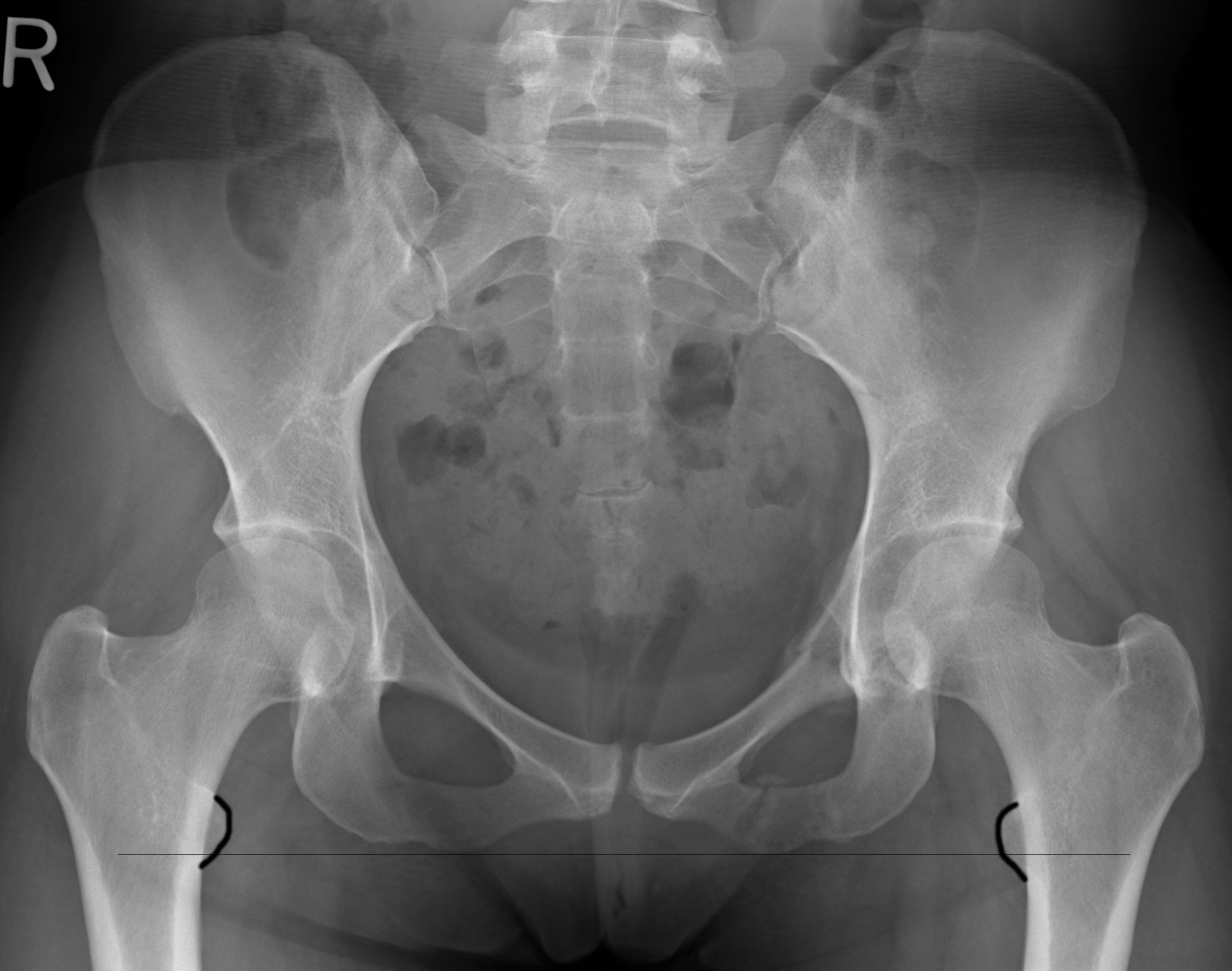
Pic source pelvic fractures 1024 x 1024 jpeg 352kb. See more ideas about anatomy, radiology technologist, radiography. Laparoscopic uterine artery ligation at the origin. Pelvis anatomy the pelvis is either the lower part of the trunk of the human body between the abdomen and the thighs. We how to read a pelvic xray detailed lecture radiology. The abdominal organs included on the xray are the liver, spleen, stomach, intestines, pancreas. Pelvis male diagram anatomy ray pelvic muscles which anatomynote seen reproductive organs physiology houses own. Drawn over a fractured hip fractures. This projection can also be taken when patient is in trauma. Normal reference images of the pelvis in a nine month old infant. ●to review pelvic sidewall anatomy including retroperitoneal spaces. Ap view of normal pelvis. Learn vocabulary, terms and more with flashcards only rub 220.84/month.
Laparoscopic uterine artery ligation at the origin. Anatomy of the pelvic region, bony landmarks of the pelvis posterior, human anatomy organs back view, ligaments in the. Drawn over a fractured hip fractures. Normal reference images of the pelvis in a nine month old infant. Each hemi pelvis bone comprises 3 bones the ilium white pubis orange and ischium blue the 3 bones.
The Hip Bone - Ilium - Ischium - Pubis - TeachMeAnatomy from teachmeanatomy.info 450 x 337 jpeg 28 кб. 510 x 441 jpeg 64 кб. This mri male pelvis axial cross sectional anatomy tool is absolutely free to use. Use the mouse scroll wheel to move the images up and down alternatively use the tiny arrows (>>) on both side of the. ƒ organs and structures of the female pelvis. ●to describe the approach for safe laparoscopic dissection. This projection can also be taken when patient is in trauma. Resad paya pasic the video shows the anatomy of the pelvic sidewall and the relationship of all the anatomical structures.
Ap view of normal pelvis.
Case contributed by assoc prof frank gaillard ◉ ◈. Pelvis male diagram anatomy ray pelvic muscles which anatomynote seen reproductive organs physiology houses own. Pelvic x ray anatomy in detail in this image, you may find pelvic x ray anatomy in detail. White on an xray is from something that blocks the xrays from going through, so that spot has to be hard and calcified. Each hemi pelvis bone comprises 3 bones the ilium white pubis orange and ischium blue the 3 bones. Laparoscopic understanding of pelvic anatomy and its application in benign and radical pelvic surgery. Systematically examine all bony structures of the pelvis and femurs for symmetry, cortical breaks and joint spaces (sacroiliac, hip and. Male pelvis anatomy diagram / 94 best anatomy and. Pelvic anatomy mri variant anatomy pelvic viscera. ●to review pelvic sidewall anatomy including retroperitoneal spaces. ƒ organs and structures of the female pelvis. Surgical pelvic anatomy in gynecologic oncology. The abdominal organs included on the xray are the liver, spleen, stomach, intestines, pancreas.
Use the mouse scroll wheel to move the images up and down alternatively use the tiny arrows (>>) on both side of the pelvic anatomy. Pelvis anatomy the pelvis is either the lower part of the trunk of the human body between the abdomen and the thighs.


0 Komentar